Figure 2.
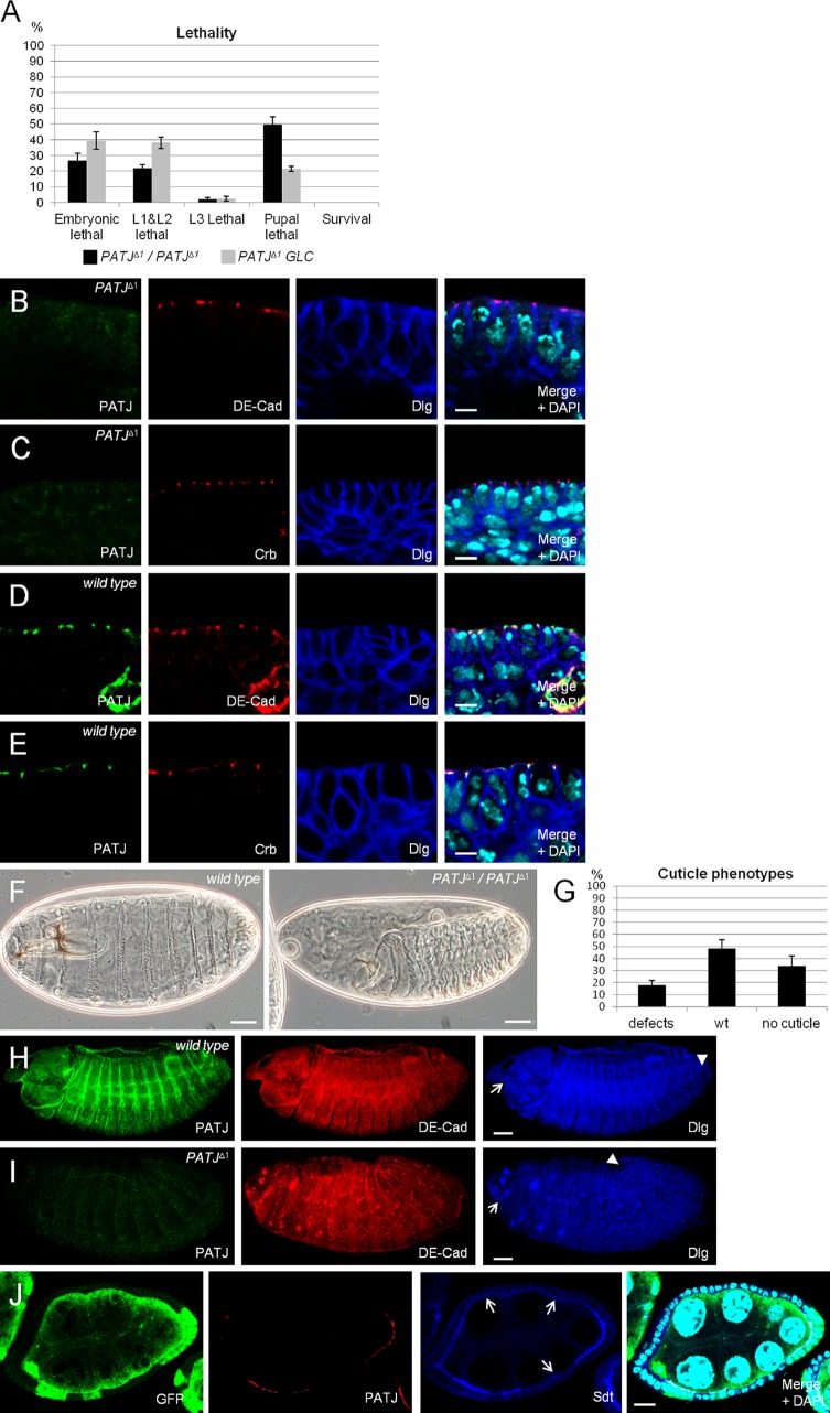
PATJ is not essential for apical–basal polarity. (A) Lethality of flies homozygous for PATJΔ1. Data were averaged from three different experiments with 100 embryos each. PATJΔ1/PATJΔ1 represent embryos homozygously mutant for PATJ that still contain the maternal component, and PATJΔ1 GLC are embryos derived from PATJΔ1 germline clones, which lack maternal and zygotic PATJ expression. (B–E) Epithelia of wild-type and PATJ mutant embryos (derived from PATJΔ1 germline clones) at stages 12/13 (shown is the mature epithelium of the embryonic epidermis), stained against DE-Cad/Dlg and Crb/Dlg, respectively. (F) Cuticle phenotypes of wild-type embryos (left) and embryos homozygous mutant for PATJΔ1 (right). (G) Quantification of cuticle phenotypes from PATJΔ1 homozygous embryos. Cuticles were scored from three independent experiments with total numbers of embryos of 174. (H and I) Overview of wild-type and mutant embryos. The head region is indicated by arrows, and the posterior end of the germband is marked by arrowheads. Note that germband retraction is not completed in the embryo homozygous for PATJΔ1, resulting in a posterior end at ∼20% embryo length. This embryo also displays head defects. (J) Follicle cell clones for PATJΔ1 showing loss of PATJ staining and decreased protein levels of Sdt at the apical junction (arrows). PATJ mutant clones are marked by the absence of GFP. wt, wild type. Error bars show SDs. Bars: (B–E) 5 µm; (F, H, and I) 200 µm; (J) 10 µm.

