
Figure 6.
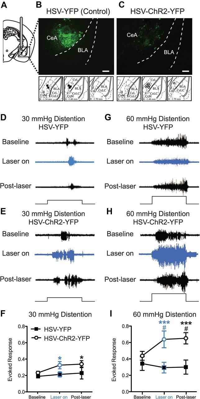
Optogenetic stimulation of right central amygdala induces bladder hyperalgesia. A, Mice are cannulated in the right CeA and HSV vectors are delivered via the cannula. B, C, YFP fluorescence (via immunohistochemistry) is seen in the CeA of both HSV-YFP (control vector) (B) and HSV-ChR2-YFP (optogenetic vector)-injected mice (C). Scale bars, 0.2 mm. Differences in staining pattern for HSV-YFP- versus HSV-ChR2-YFP-treated mice are likely due to the fact that there is probably higher expression of cytoplasmic YFP (i.e., HSV-YFP) compared with the ChR2-YFP fusion protein, which is membrane bound. Brain atlas insets show cannula targeting with black squares indicative of cannula tips of HSV-YFP mice and white hexagons indicative of cannula tips of HSV-ChR2-YFP mice. Representative images of raw EMG traces from HSV-YFP- or HSV-ChR2-YFP-injected mice during 30 mmHg (D, E) and 60 mmHg (G, H) bladder distention. Optic stimulation of HSV-ChR2-YFP-treated mice (n = 7) increases the evoked response to both 30 and 60 mmHg bladder distention compared with baseline (prestimulation) responses (E, H) and increases the evoked response to 60 mmHg bladder distention compared with HSV-YFP-injected mice (n = 5) (I). Laser light alone does not increase the evoked response to bladder distention in HSV-YFP mice (D, F, G, I). The black steps in traces show 20 s bladder distention stimuli (*p < 0.05; ***p < 0.001, Bonferroni's post test compared with baseline for HSV-ChR2-YFP mice; #p < 0.05, Bonferroni's post test compared with HSV-YFP control mice at indicated time points). Error bars indicate SEM.