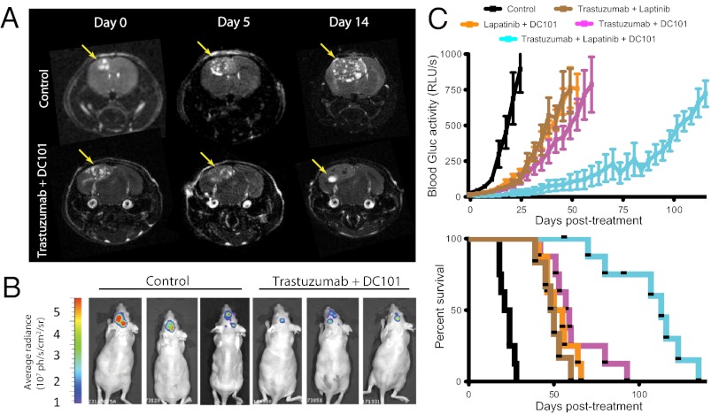
Fig. P1.
Effect of various combinations of anti-HER2 and anti-VEGFR2 treatments on the growth of established breast cancer brain metastases. Breast cancers were allowed to grow to roughly 10 mm3 in size in the brain parenchyma before the initiation of treatment, which consisted of either a dual or triple combination of anti-HER2 agents (trastuzumab and lapatinib) and an anti-VEGFR2 antibody (DC101). (A) MRI scans of control and the combination treatment of trastuzumab and DC101 at days 0, 5, and 14. Arrows indicate tumor location. (B) Bioluminescence images of control- and trastuzumab plus DC101-treated mice at day 14. ph, photons; sr, steradian. (C) Tumor growth plot (Upper, mean ± SEM) and Kaplan–Meier survival plot (Lower) of tumor-bearing mice treated with control (black); trastuzumab plus DC101 (magenta); lapatinib plus DC101 (orange); trastuzumab plus lapatinib (brown); and the triple combination of trastuzumab, lapatinib, and DC101 (cyan) (n = 6–9 mice). RLU/s, Relative Light Units per second.

