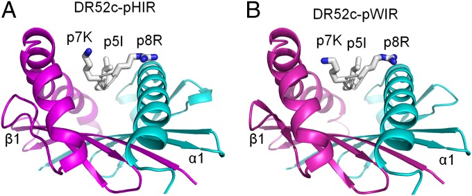
Fig. 3.
The side chains of critical mimotope amino acids p5I, p7K, and p8R are surface-exposed on the mimotope–DR52c complexes. The structures of two of the mimotopes in Fig. 1C, (A) pHIR and (B) pWIR, bound to DR52c were solved to resolutions of 2.2 and 2.5 Å, respectively. The peptide binding grooves are viewed from the C terminus of the peptides. Ribbon representations of the α1 (cyan) and β1 (magenta) DR52c helices and the peptide backbone (white) are shown with wire-frame representations of surface-exposed side chains of p5I, p7K, and p8R [CPK (Corey, Pauling, Koltun) coloring].

