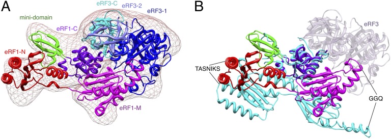
Fig. 2.
Structural dynamics of the eRF1–eRF3 complex. (A) Conformation of the eRF1–eRF3–GMPPNP ternary complex when bound to the pretermination 80S ribosome. Individual domains for each factor are labeled and colored separately. Models of eRF1 and eRF3 determined by X-ray crystallography were docked into the cryo-EM map. (B) Superimposition of human apo-eRF1 (cyan) with that of the observed eRF1 structure in the 80S–P-tRNA–eRF1–eRF3–GMPPNP complex. The latter is labeled and color coded by domain. Alignment of the two structures was facilitated by superimposing the C-terminal domain of eRF1. The position of eRF3 in the ternary complex is shown in transparent blue.

