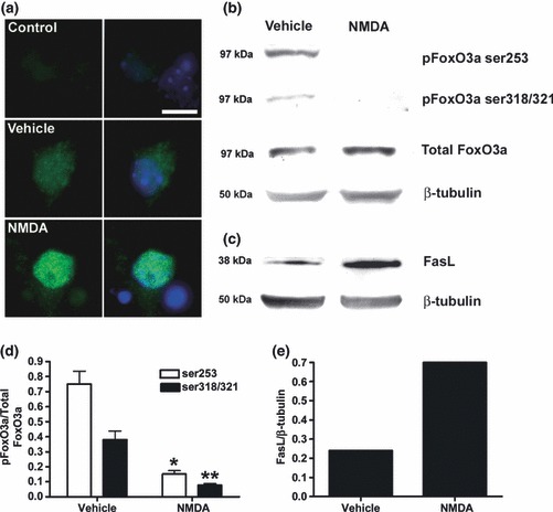
Fig 1.

NMDA inhibits FoxO3a phosphorylation, stimulates FoxO3a nuclear translocation and up-regulates FasL. Cortical neurones were treated with NMDA (50 μM) for 2 h, and FoxO3a phosphorylation and FoxO3a translocation measured by immunoblotting (b, d) and immunocytochemistry, respectively, (a) and FasL expression by immunoblotting only (c, e). Reduced dephosphorylation of both pFoxO3a ser253 and pFoxO3a ser319/321 was evident following exposure to NMDA (b, d) with no change in total levels of FoxO3a or β-tubulin, but was associated with an increase in the levels of FasL (c, e). Blots shown are representative of three independent experiments. (d) Data presented as mean band intensity (mean ± SEM, n = 3) expressed as pFoxO3a relative to total FoxO3a following exposure to Vehicle control or NMDA (*p < 0.05, **p < 0.01). (e) Data presented as mean band intensity (mean ± SEM, n = 3) expressed as FasL relative to β-tubulin following exposure to Vehicle control or NMDA. To determine whether NMDA-mediated dephosphorylation of FoxO3a resulted in the nuclear accumulation of FoxO3a, cells were probed with an anti-FoxO3a antibody [(a), left and right panels] and counter-stained with Hoe 33342 (right panel only) to label nuclei. Control represents staining because of secondary antibody only. Under basal conditions, FoxO3a was distributed throughout the cytoplasm of cortical neurones with some diffuse staining evident in the nucleus (a, Vehicle). Following treatment with NMDA, FoxO3a staining was almost exclusively nuclear (a, NMDA). Scale bar = 20 μm.
