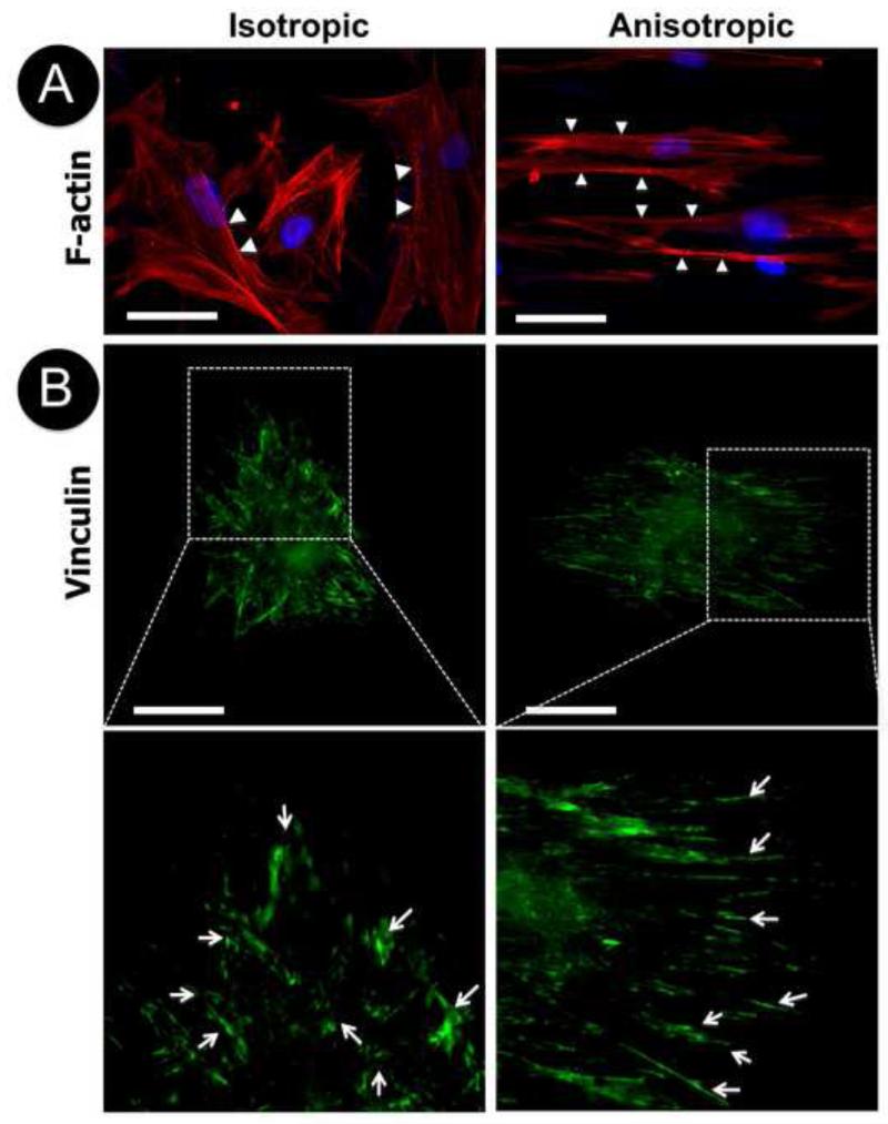
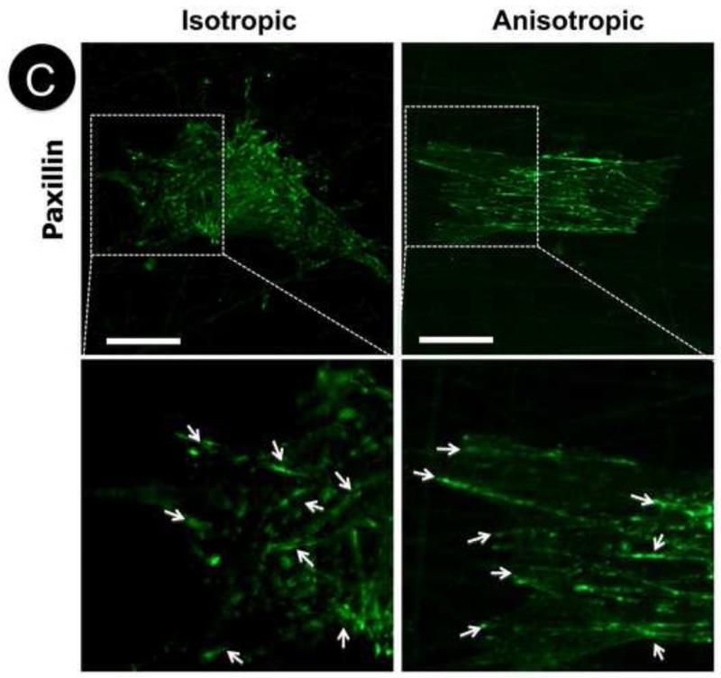
Figure 2.
Cellular morphology of human dermal fibroblasts on isotropic and anisotropic PCL/Coll nanofibrous matrices. Immunofluorescent staining of intracellular cytoskeleton protein of F-actin (red/orange) and nuclei with DAPI (purple/blue) (A), and focal adhesion proteins of vinculin (green) (B) and paxillin (green) (C). White arrows indicate spatial distribution of F-actin, vinculin or paxillin. Scale bar: 50μm.


