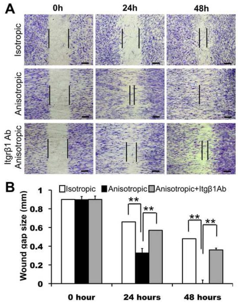
Figure 6.
Migration of human dermal fibroblasts on PCL/Coll nanofibrous matrices in wound healing assay. (A) Cells (5×10⁴ per matrix) were seeded on either isotropic or anisotropic matrices with an insert in the middle. After 12 hours, the insert was removed to generate a 0.9-mm “wound gap”. Cells were allowed to migrate into the wound gap, and visualized after 24 and 48 hours using methylene blue staining. (B) Quantification of the “wound gap” distance between the front lines of migrating fibroblasts (n=4). ** Statistically significant, p <0.01. Scale bar: 300μm

