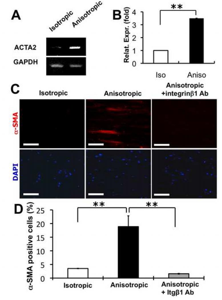
Figure 7.
Myofibroblastic differentiation of human dermal fibroblasts on PCL/Coll nanofibrous matrices. (A) RT-PCR and (B) Q-PCR analyses of cellular α-SMA (ACTA2) gene expression level on either isotropic or anisotropic nanofibrous matrices. (C) Immunostaining of cellular α-SMA proteins on isotropic and anisotropic matrices. (D) Quantification of α-SMA positive cells. The DAPI stained cell nuclei were used for normalization. ** Statistically significant, p <0.01. Scale bar: 100 μm.

