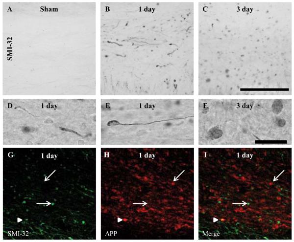
Figure 4.
Intra-axonal accumulation of dephosphorylated 200-kDa neurofilament subunit following diffuse brain injury in the immature rat. (A-F) Representative photomicrographs of SMI-32-labeled axons within the corpus callosum of sham (A), and brain-injured rats at 1 (B) and 3 days (C) post-injury. (D) An example of SMI-32-labeled swollen contiguous axons. (E, F) Examples of terminal bulbs. (G-I) An example of double-label immunofluorescence for intra-axonal amyloid precursor protein (APP) accumulation (red) and SMI-32 immunoreactivity (green) at 1 day post-injury. Single-labeled profiles (either APP-positive/SMI-32-negative or APP-negative/SMI-32-positive) are denoted by arrows; double-labeled profiles (APP-positive/SMI-32-positive) are denoted by arrowheads. Photomicrographs were obtained at 63x magnification. Scale bars in panels C (20x) and F (100x) represent 100 μm for panels A-C and D-F, respectively.

