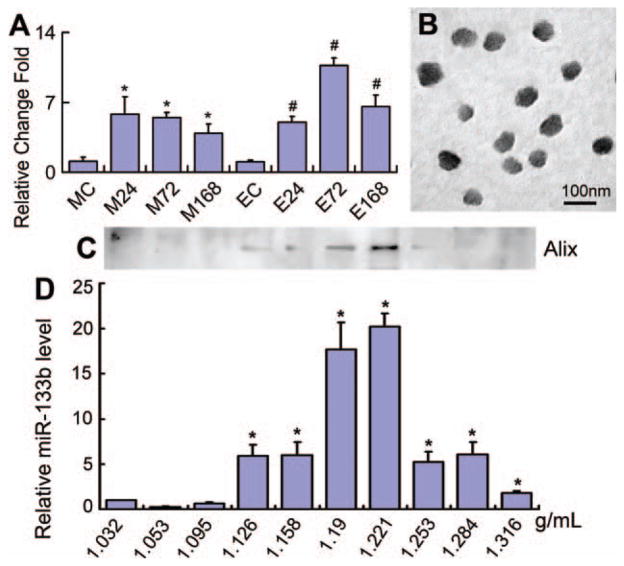
Figure 2.
Culture of mesenchymal stromal cells (MSCs) with ischemic tissue brain extracts increased the miR-133b expression in MSCs and their generated exosomes. Real-time reverse-transcribed PCR data showed that compared to the normal rat brain tissue extract treated group, miR-133b levels in MSCs exposed to middle cerebral artery occlusion (MCAo) brain tissue extracts and their exosomes were significantly increased, and the exosome miR-133b level reached a peak after MSCs cultured with the 72 hours post-MCAo brain tissue extract (A). The TEM image showed that the morphology of MSC released exosomes within a size range of 40–100 nm (B). Western blot detected that the exosome marker protein, Alix, was primary located in the range of density from 1.19 to 1.221 g/ml (C), and miR-133b also primary presented a high level in these two density fractions (D). MC, EC: MSCs and their exosomes exposed to the normal rat brain tissue extract as controls; M24, M72, M168, E24, E72, and E168: MSCs and their exosomes treated with the brain tissue extracts after MCAo at 24, 72, and 168 hours, respectively. *, p < .05, compared with MC in (A) and compared with 1.032 g/ml density fraction in (D). #, p < .05 compared with EC (n = 3 per group). Abbreviation: miR-133b, microRNA 133b.

