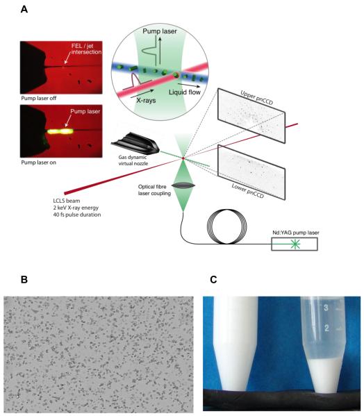
Figure 1.
(a) Experimental setup for serial femtosecond crystallography. Tiny crystals are injected into an X-FEL beam using a liquid microjet. This setup is very convenient for pump-probe experiments using laser excitation. Shown is an experiment on the photo-induced dissociation of photosystem I-ferredoxin cocrystals, taking place on a microsecond time-scale (from ref [43]). (b) Microcrystalline sample for serial femtosecond crystallography data collection. Lysozyme crystals (≤1 × 1 × 3 μm3) as seen through a conventional light microscope, (c) Microcrystalline lysozyme suspended in solution (left) and after settling (right) (from ref.[42]). It is important to note that the quantities depicted here are by no means excessive; several milliliters of very dense crystalline slurry are currently required for SFX experiments.

