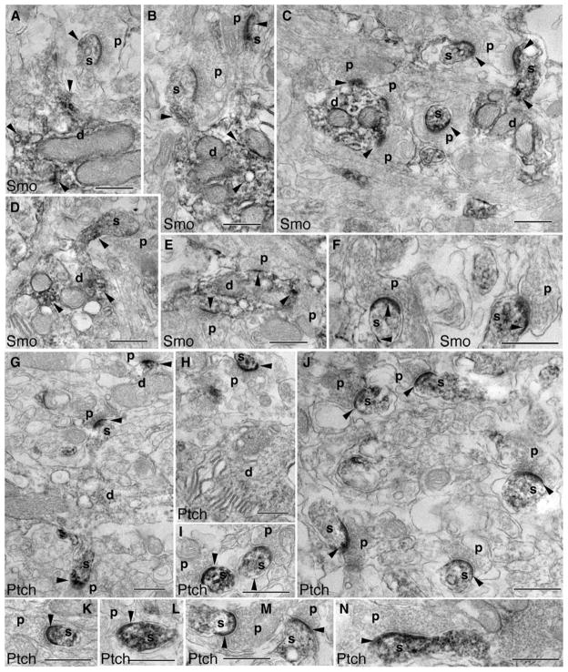
Fig. 4.
Subcellular distribution of Smo and Ptch1 in the molecular layer of the adult cerebellum, revealed by immunoperoxidase/DAB electron microscopy. a–f Prominent Smo labeling (arrowheads) in dendrites (d) and postsynaptic spines (s) of Purkinje cells. Smo-labeled spines are commonly opposed to unlabeled presynaptic terminals (p) of parallel fibers. Postsynaptic Smo labeling is also seen in synapses on the dendrite shaft of interneurons (c, e). g–n Examples of Ptch1 labeling. Compared to Smo, Ptch1 labeling (arrowheads) is usually not prominent in dendrites (d) but is dense in postsynaptic Purkinje spines (s). Dendrites (d) shown in g (bottom) and h are from Purkinje cells, and another in g (top) is from an interneuron. Ptch1-labeled Purkinje cell spines are opposed to unlabeled presynaptic terminals (p) from parallel fibers (g–m); postsynaptic labeling also is seen in some dendrite shaft synapses (g, top). n A climbing fiber synapse with prominent postsynaptic Ptch1 labeling. Scale bars, 500 nm

