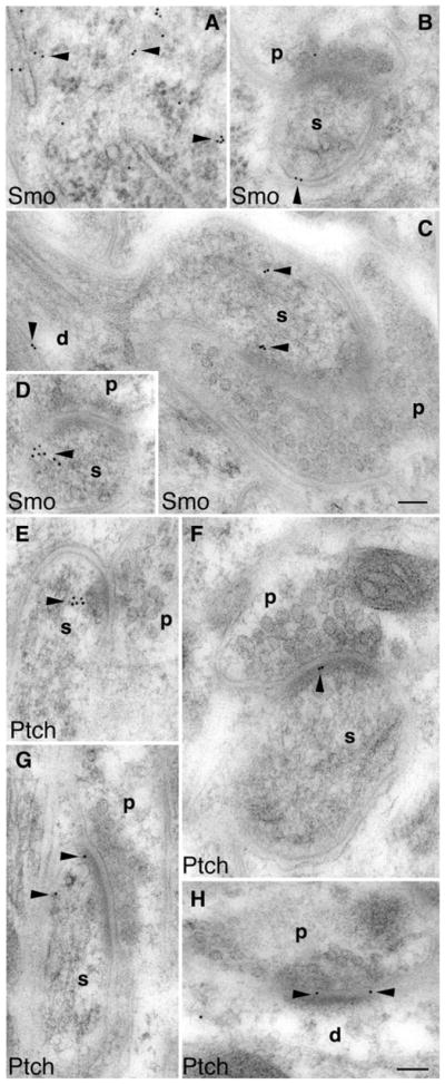
Fig. 5.

Immunogold electron microscopy also shows postsynaptic localization of Smo and Ptch1 in the molecular layer of the adult cerebellum. a Smo labeling (arrowheads) in the cell body of a Purkinje cell. b and d Smo labeling in postsynaptic Purkinje spines (s) opposed to the presynaptic terminals (p) of parallel fibers. c Smo labeling in a Purkinje dendrite (d) as well as its spine (s) that is opposed to a presynaptic terminal (p) of a climbing fiber. e–g Ptch1 labeling (arrowheads) in postsynaptic Purkinje spines (s) opposed to the presynaptic terminals (p) of parallel fibers. h Postsynaptic Ptch1 labeling at a dendritic (d) synapse of an interneuron. Scale bars, 100 nm
