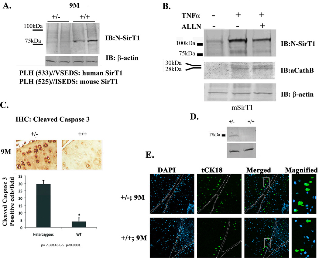
Figure 2. 9M SirT1+/− mice show enhanced cleaved caspase 3 as compared to WT.
A. Protein extracts from duplicate SirT1 +/− vs. duplicate WT mice were obtained and immunoblotted with an N-SirT1 antibody. Lower panel shows the predicted cleavage site for human and mouse SirT1, which generate a similar molecular weight cleaved SirT1 product. B. Stably transfected human chondrocytes expressing mSirT1 were treated as indicated above the blot and expressed enhanced 75SirT1 and active Cathepsin B (aCathB) in the presence of TNFα. C. Immunohistochemistry of the MTP at x20 magnification (upper panel) and equivalent densitometry for chondrocytes positive for cleaved caspase 3 in adult SirT1 +/− and WT mice. D. Immunoblot analyses for cleaved caspase 3 in adult SirT1 +/− vs. WT mice. A.U denotes arbitrary units of intensity. E. shows increased truncated cytokeratin 18 (tCK18) pronounced in SirT1+/− (2-fold) vs. WT. mice. DAPI was used to stain the nuclei and white bars differentiate between the medial tibial plateau (left) and the medial femoral plateau (left). Bars represent a mean and standard error of n=6 (with 2 repetitions per sample). Protein extracts were obtained from 3 different mice or 3 different stable cell lines expressing mSirT1.

