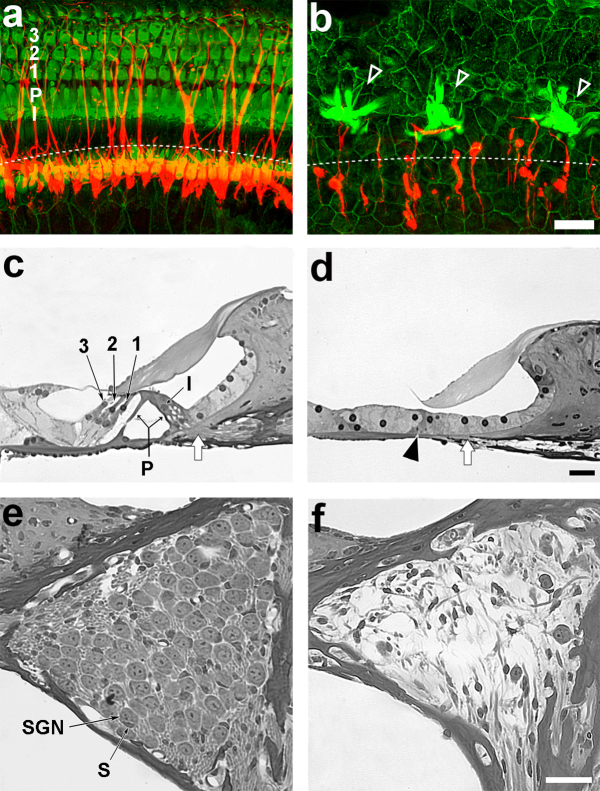
Figure 1. The auditory epithelium in wild-type mouse ears (a, c and e) or Pou4f3 homozygous mutant ears (b, d and f) stained for neurofilaments (red) and actin (green) and viewed by epi-fluorescence (a, b) or light microscopy of cross-sections (c-f).
All specimens were obtained from 6 week old mice. The left (lateral) side of the cross-section corresponds to the upper (lateral) aspect of the whole-mounts. (a) The actin staining shows the epithelial mosaic containing hair cells and supporting cells. The neurofilament-positive nerve fibers extend from modiolar aspect to the inner and outer hair cells. The approximate site of the habenula perforata (HP) is depicted by a dotted line. (I, inner hair cell; P, pillar cells; 1, 2, 3, 1st 2nd and 3rd rows of outer hair cells). (b) The Pou4f3 mutant ears have no sensory hair cells and the disorganized junctional contour resembles a flat epithelium. Actin rich (AR) cells are clustered in patches along the auditory epithelium (open arrow-head). A few nerve fibers are seen and appear to preferentially grow toward the AR cells, but not beyond them. The dotted lines show the area of the HP. (c) The normal organ of Corti contains a highly organized sensory epithelium with inner and outer hair cells and non-sensory cells. The inner and outer pillar cells (P and arrows) flank the tunnel of Corti. The site of the HP is depicted with a white arrow. (d) In Pou4f3 mutants, the auditory epithelium resembles a flat epithelium, composed of a single layer of cuboidal epithelial cells and absence of the tunnel of Corti (arrow-head). The presumed site of the HP is depicted with a white arrow. (e) Rosenthal's canal contains densely packed SGNs and a dense array of myelinated fibers in a wild-type ear (SGN, spiral ganglion neuron; S, Schwann cell). (f) The SGNs appear smaller, and the density of SGNs is reduced in Pou4f3 mutant ear. All scale bars indicate 20 µm.

