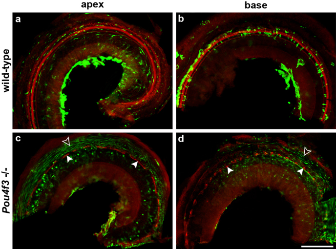
Figure 3. Low magnification epi-fluorescence images showing the distribution of GFP in wild-type (a, b) or Pou4f3 homozygotes (Pou4f3 −/−) cochleae (c-d) following inoculation of Ad.eGFP.
Whole-mounts are double-stained with phalloidin (red) and GFP (green) and show cochlear apex (a and c) and base (b and d). (a) GFP can be detected in numerous mesothelial cells (elongated or spindle shaped cells) throughout the entire cochlear apex. (b) Expression of GFP is seen in several types of cells including supporting cells and inner hair cells, as well as in mesothelial cells. (c, d) In Pou4f3 mutants, GFP expression is detected in a large number of mesothelial cells (open arrow heads), but not in AR cells (filled arrow heads). Scale bar indicates 100 µm.

