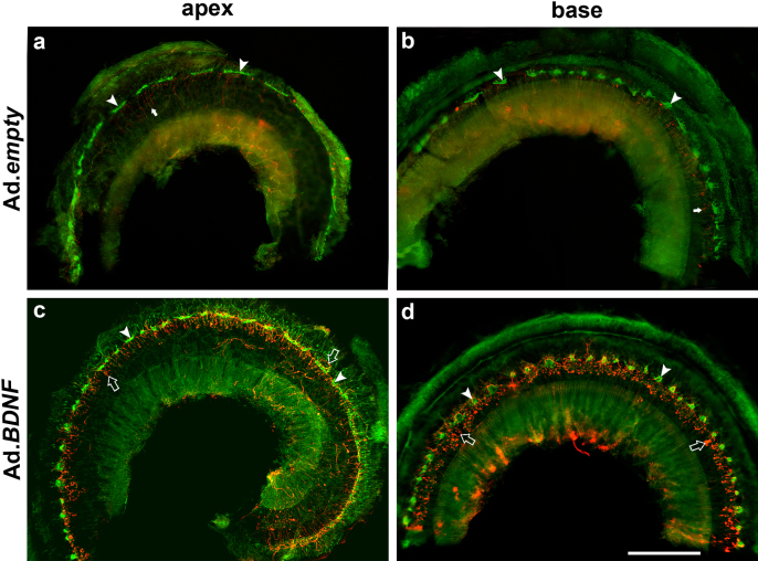
Figure 4. Whole-mounts of Pou4f3 mutant cochleae treated with Ad.empty (a, b) or Ad.BDNF (c, d), stained for actin (green) and neurofilament (red) and imaged with epi-fluorescence (Figures a and c for apex, b and d for base).
(a, b) After Ad.empty inoculation, there is no visible effect on AR cells or on nerve fiber distribution throughout the cochleae (arrows). AR cells are evenly spaced along the cochlear duct (arrow heads). (c, d) Following Ad.BDNF inoculation, many nerve fibers are observed around AR cells (open arrows) throughout the cochlear duct, as seen in both the apex (c) and the base (d). The shapes of AR cells in Ad.empty cochleae are similar to that seen in Ad.BDNF-treated ears (arrow heads). Scale bar indicates 100 µm.

