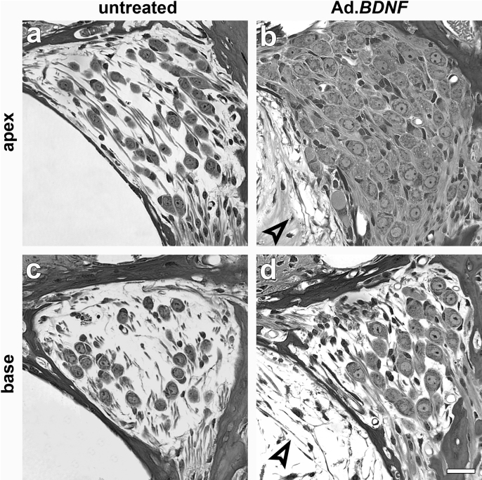
Figure 7. Light microscope micrographs of cross-sections through Rosenthal's canal in the apex (a-b) and base (c-d) of contralateral ears (a and c), and 2 weeks after inoculation of Ad.BDNF (b and d).
(a) SGNs are much less dense than normal in the apical cochlear turns of untreated ears. (b) Rosenthal's canal of the treated ear appears densely packed with SGNs and morphologically healthy, with normal appearing nuclei and cytoplasm. (c) SGNs in the base appear similar to those seen in the apex in the absence of BDNF treatment (a). (d) BDNF-induced preservation of SGNs in the base is less complete than in the apex (compare to (b). In some of the Ad.BDNF-inoculated ears, connective tissue could be observed in the scala tympani (open arrow-heads in b and d). Scale bar indicates 20 µm.

