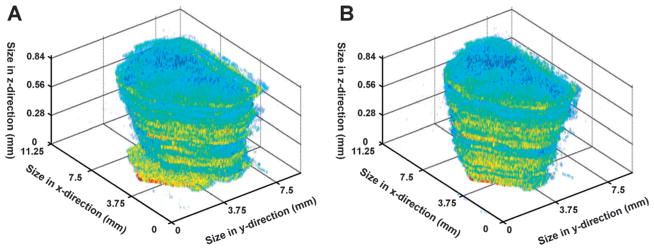
Fig. 2. Section alignment of acquired 2D images.
(A) An unaligned 3D image of intensities for an ion at m/z 97 demonstrates the small but significant shifts in x and y and the slight rotational variation of the x-y plane when comparing sequential sections. (B) Following the automated alignment strategy, sequential sections in 3D appear to be highly correlated along the z-axis.

