
Fig. 6. Cut-away view of 3D tumor model.
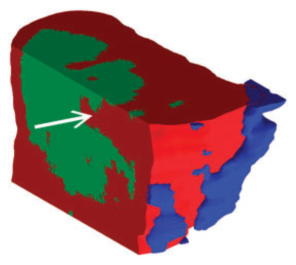
The reference viewpoint can be set to any arbitrary position and cut-aways of any planar surface allow inspection of the interior of the tumor. By looking at depth profiles along dimensions other than the z-axis, one can see how various ions are distributed in micro-encapsulated pockets (marked by white arrow) which might remain undetected when looking only at single-sections.