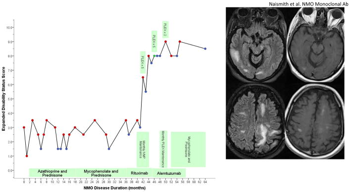
Figure.
Timeline of clinical events and treatments with Expanded Disability Status Scale. Relapses treated with IVMP are shown by red dots, relapses treated with PLEX are noted (red dots are IVMP, blue dots were non-relapse exam visits, green dot is PLEX without IVMP). After alemtuzumab, brain MRI revealed multiple, large regions of T2 signal abnormality with patchy enhancement. This included a 1.5 × 1.5 × 4 cm lesion in the right temporal lobe, a 1.4 × 1.8 × 1.2 cm lesion in the right corona radiata, and a 5.5 × 2.5 × 2.4 cm lesion in the left occipital lobe.

