Figure 3.
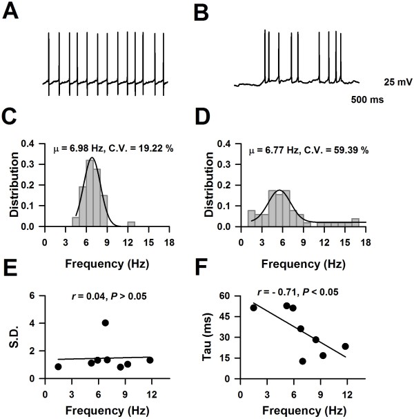
Firing patterns of neurons in LI of the ACC from adult mice. A), The representative showed one example of a spontaneous firing neuron in the LI of the ACC. B), The representative showed one example of d-AC neurons with fAHP in the LI of the ACC. C), The representative showed one example of d-AC neurons with sAHP in the LI of the ACC. Ca, Under subthreshold current stimulation, the membrane potential of this neuron developed a ramp depolarization. D &E), The representatives showed examples of neurons with ADP, named ADP1 and ADP2, respectively. a, the representative traces showed the change of membrane potential under the currents stimulation, the intensity of the currents were around the Rheobase of this neuron. b, The inset showed the AHP characteristics of the recorded neuron. c, Example traces showed the membrane potential change under currents stimulation. The APs fired at maximal firing frequency (for the definition, see the method part). d, the firing frequency changed under current stimulation, data was fitted by function: . (For detail, see method part).

