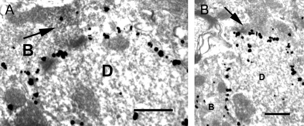
Figure 8.
Electron micrographs showing axo-dendritic contacts between P2X3-positive boutons (arrows) and NK-1r-positive dendrites (D) (A and B). Note the silver-intensified gold particles along the plasma membrane of the dendrites, representing NK-1r-IR sites. B, unlabeled axonal bouton. Scale bar = 0.5 μm.

