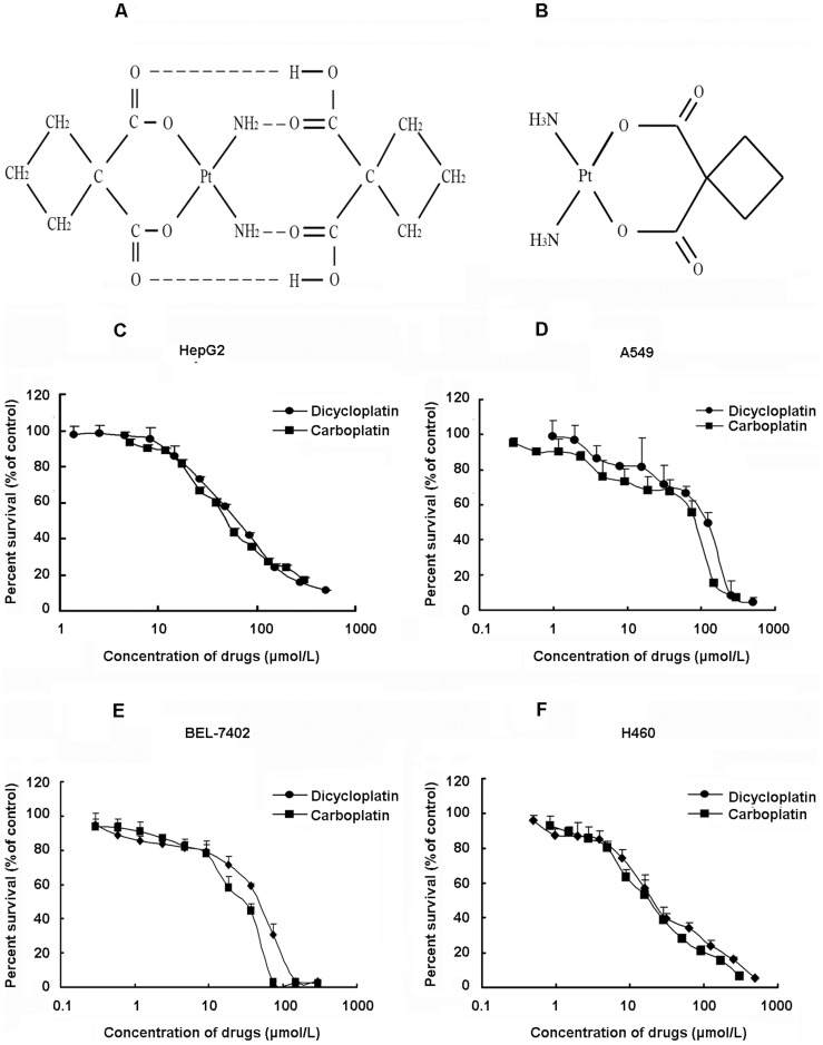
Figure 1. Cytotoxic effect of dicycloplatin in cancer cell lines.
A, structure of dicycloplatin. B, structure of carboplatin. C–F, cytotoxicity of dicycloplatin and carboplatin in HepG2, A549, BEL-7402 and H460 cells. Cytotoxicity was measured by MTT assay. The cells were exposed to a full range of concentrations of dicycloplatin and carboplatin for 72 h. Cell viability with a model 550 microplate reader after staining with MTT for 4 h. The data presented represent mean±SD of three independent experiments.

