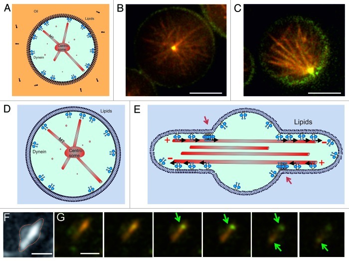
Figure 4. Dynein-mediated centrosome positioning in emulsion droplets and liposomes. (A–C) Centrosome positioning in emulsion droplets. (A) Cartoon of the experiment. Dynein molecules are linked to phospholipids at the surface of the droplet. (B–C) Preliminary experiments show that dynein molecules attached to phospholipids can either center (B) or decenter (C) asters. MTs (red) growing from a purified centrosome interact with dynein (green) at the edge of the droplets. Shown is a single Z-plane of a spinning disk confocal fluorescence stack. Scale bars: 10 µm. (D) Centrosome positioning in GUVs. Cartoon of the desired experiment. (E) Cartoon explaining the accumulation of dynein at the entrance of the protrusions created by free MTs. Red arrows point to the accumulations. (F–G) Free taxol-stabilized MTs grown in GUVs, with membrane-bound dynein molecules. Scale bar: 3 µm (F) Z-projection of fluorescent MTs. (G) Individual Z-planes of the GUV seen in (F). Shown is a superposition of the MT (red) and dynein (green) signals. Arrows indicate the positions of dynein accumulation at the entrance of the protrusions. Z spacing = 0.3 μm. Scale bar: 3 µm.

An official website of the United States government
Here's how you know
Official websites use .gov
A
.gov website belongs to an official
government organization in the United States.
Secure .gov websites use HTTPS
A lock (
) or https:// means you've safely
connected to the .gov website. Share sensitive
information only on official, secure websites.
