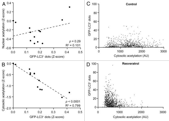
Figure 5. Negative correlation between the acetylation of cytoplasmic proteins and autophagy induction. GFP-LC3-expressing human osteosarcoma U2OS cells were left untreated or treated with 30 µM of the phenolic compounds used in this study, including resveratrol, for 6 h. Thereafter, cells were processed for the immunofluorescence microscopy-assisted quantification of GFP-LC3+ dots and nuclear (A) or cytoplasmic (B–D) protein acetylation levels. In (A and B), population-based results (each dot representing the mean of n = 500 cells) are reported, together with linear regression curves and the corresponding statistical indicators (p values and determination coefficients, R2). In (C and D), results from one representative experiment are reported (each dot representing a single cell). In this latter scenario, p values and determination coefficients were invariably were > 0.2 and < 0.1, respectively.

An official website of the United States government
Here's how you know
Official websites use .gov
A
.gov website belongs to an official
government organization in the United States.
Secure .gov websites use HTTPS
A lock (
) or https:// means you've safely
connected to the .gov website. Share sensitive
information only on official, secure websites.
