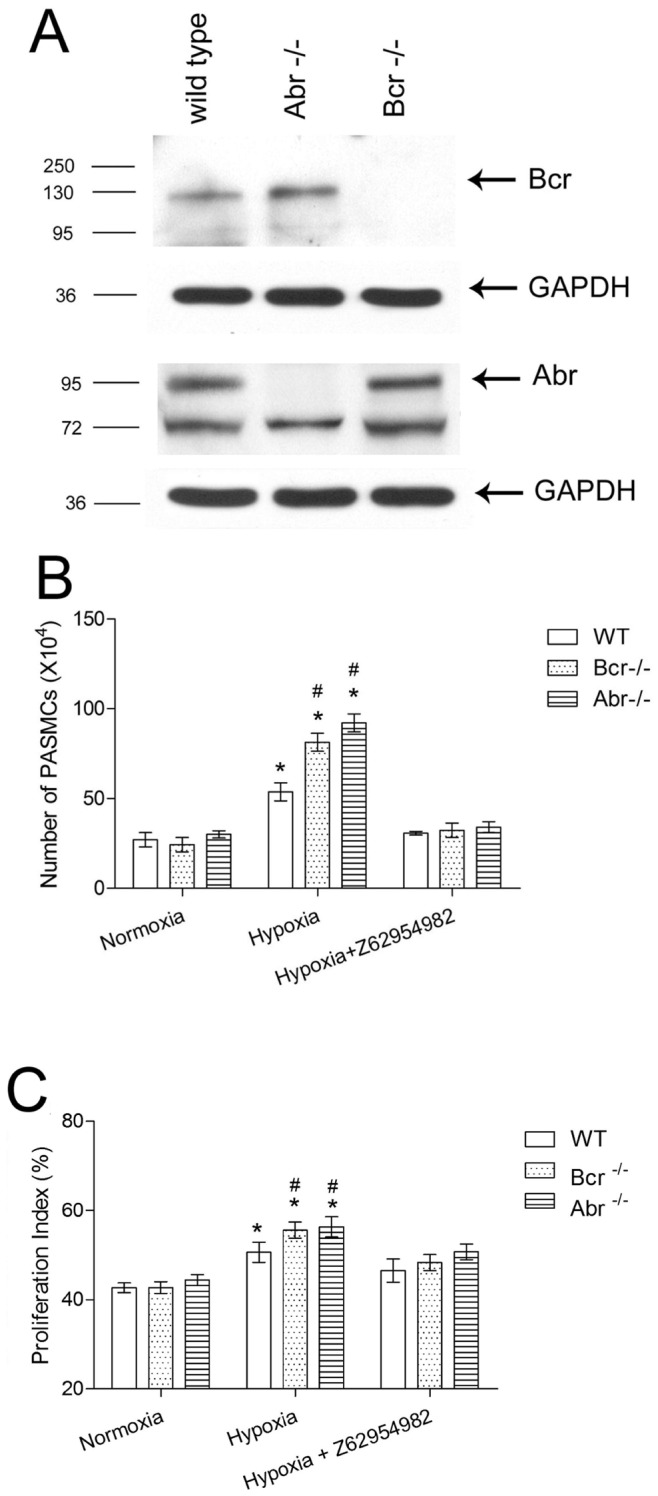
Figure 4. Bcr−/− and abr−/− PASMC show increased proliferation when exposed to hypoxia in vitro. A,

Western blot analysis on PASMC lysates from wt, bcr−/− or abr−/− mice with anti-Bcr N20 antibodies or Abr antiserum. GAPDH, loading control. B, Third passage primary PASMC isolated from the intrapulmonary arteries of 5 different mice per genotype (1×10⁴ cells/well) were synchronized by serum free medium for 24 hrs, then cultured in medium with 10% FBS for 5 days, after which cells were counted. The Rac inhibitor Z62954982 was added to the indicated samples. * p<0.05 compared with the outcomes from the same genotype PASMCs in normoxia. # p<0.05 compared with wt PASMCs exposed to the same condition. Bars, mean ±SD of triplicate wells. C, Proliferation index of wt, bcr−/− and abr−/− PASMCs was calculated as described in Methods. Flow cytometry analysis was done based on 10,000 PASMCs per sample, 3 samples per genotype per condition. * p<0.05 compared with the outcomes from the same genotype PASMCs in normoxia. # p<0.05 compared with wt PASMCs exposed to the same condition. Bars are shown as mean ±SD of triplicate wells.
