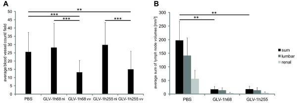
Figure 5.
Analysis of lumbar and renal lymph node metastases in PC-3 tumor-bearing mice and of Here, the blood vessel density should be mentioned first (Figure5A, text labeled in yellow) and lymph nodes second (Figure5B). (A) Blood vessel density in PC-3 tumor sections, 7 days p.i. For GLV-1h68 and GLV-1h255 blood vessels in infected (vv) and non-infected (ni) areas were counted. Infected areas were determined by expression of the viral marker GFP. For each image (200x, Leica MZ16 FA) blood vessels crossing 5 equidistant lines were counted. (B) Analysis of renal and lumbar lymph node enlargement. PC-3 tumor-bearing mice were injected i.v. with GLV-1h68, GLV-1h255 or PBS. The mice were sacrificed 24 days p.i. and lymph nodes were measured (n = 6–7).

