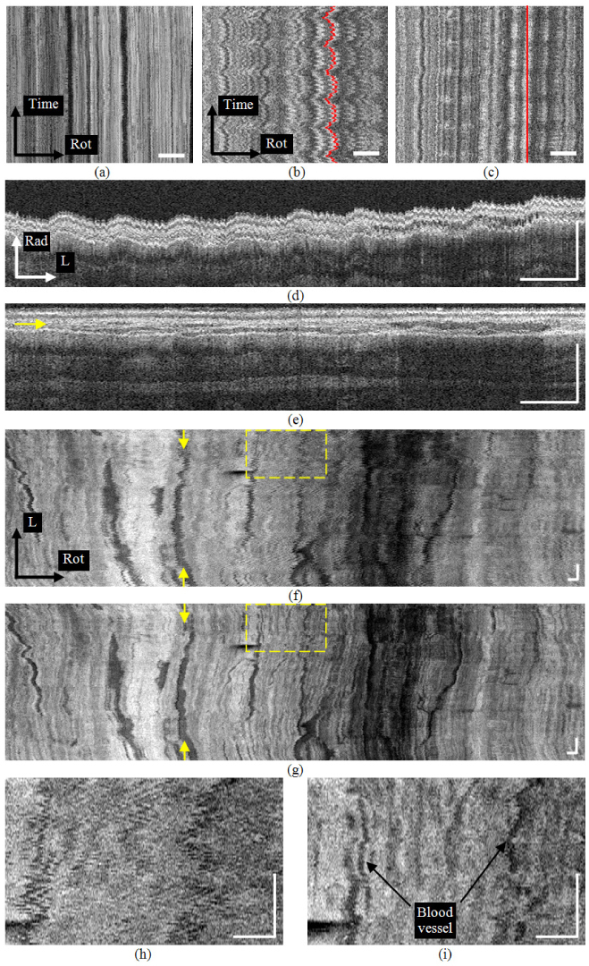
Fig. 4.

(a) The en face view of the muscularis mucosa layer in the excised esophagus from a 2-D time sequence of images. (b) The en face view of the in vivo 2-D sequence where physiological motion artifact is prominent. (c) The registered en face view of (b). (d) A radial-longitudinal view from the original 3-D sequence in vivo. (e) The registered view of (d). The arrow indicates the position of (g). (f) The en face view from the original 3-D sequence. Arrows indicate the position of (d). (g) The registered view of (f). Arrows indicate the position of (e). (h) The enlarged view of the square in (f). (i) The enlarged view of the square in (g). (Rad: radial; L:longitudinal; Rot:rotational. Bars: 1mm)
