
Fig. 5.
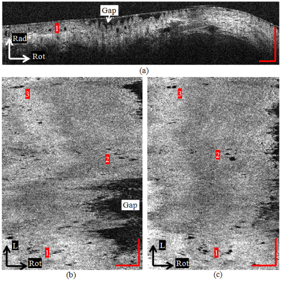
(a) A cross-sectional image of BE with high-grade dysplasia. Dark oval glandular structures can be clearly observed (example indicated by number 1). (b) The en face view at 300 μm deep within the dysplastic esophagus before rotational motion artifact correction. The shape and number of glands are significantly distorted. (c) The en face view after rotational correction. The registration algorithm more clearly reveals the glandular morphology