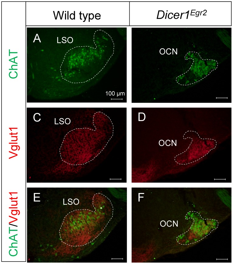
Figure 5. Presence of the olivocochlear neurons in Dicer1Egr2 mice.
A,B ChAT immunolabeling was detected throughout the LSO of wild type mice (A), whereas in Dicer1Egr2 mice, ChAT positive cells were restricted to a densely packed ventrolateral cell group (B). (C–F) The same SOC area was labeled by Vglut1, as revealed in the overlay (E–F). Two animals aged P15–P20 were analyzed per genotype. LSO, lateral superior olive; OCN, olivocochlear neurons. Dorsal is up, lateral is to the right.

