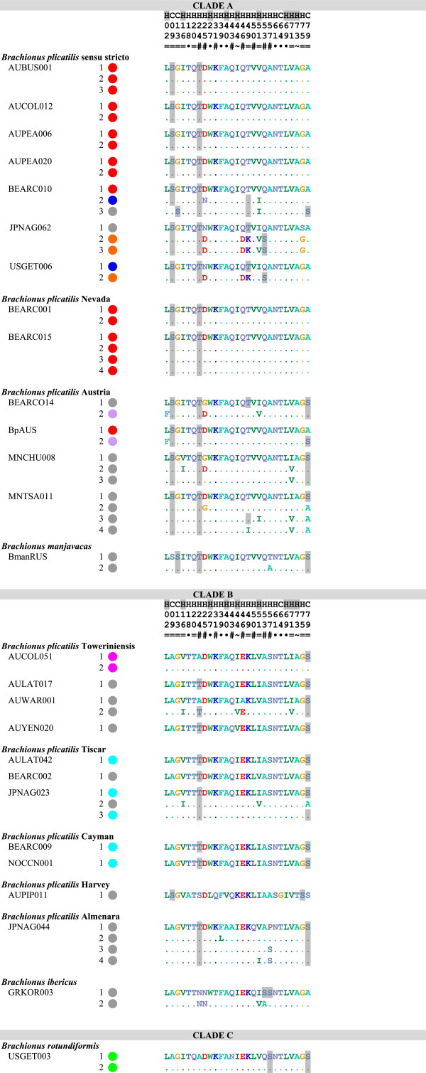
Figure 2.
Amino acid identity among terminal repeat motifs of MMR-B. All gene copies are listed for each species. Each circle represents the number of repeat units in each gene copy; colors indicate identity of repeats. Grey represents repeats with unique amino acid sequences, all other colors represent repeats found multiple times within Clade A or Clade B. Numbered polymorphic positions are shown to the right, with differences from the first repeat of the shortest gene copy shown for each isolate; predicted phosphorylated amino acids are shaded. The consensus prediction of the position being part of a coil (C) or helix (H) is shown above the position numbers, with positions predicted to be buried in the hydrophobic core shaded. Symbols below the position numbers indicate if the polymorphism is unique to a single repeat (·), alternates between only two (=) or almost always two (~) residues, or is more polymorphic (#). Underlining indicates synapomorphies between Clades A and B.

