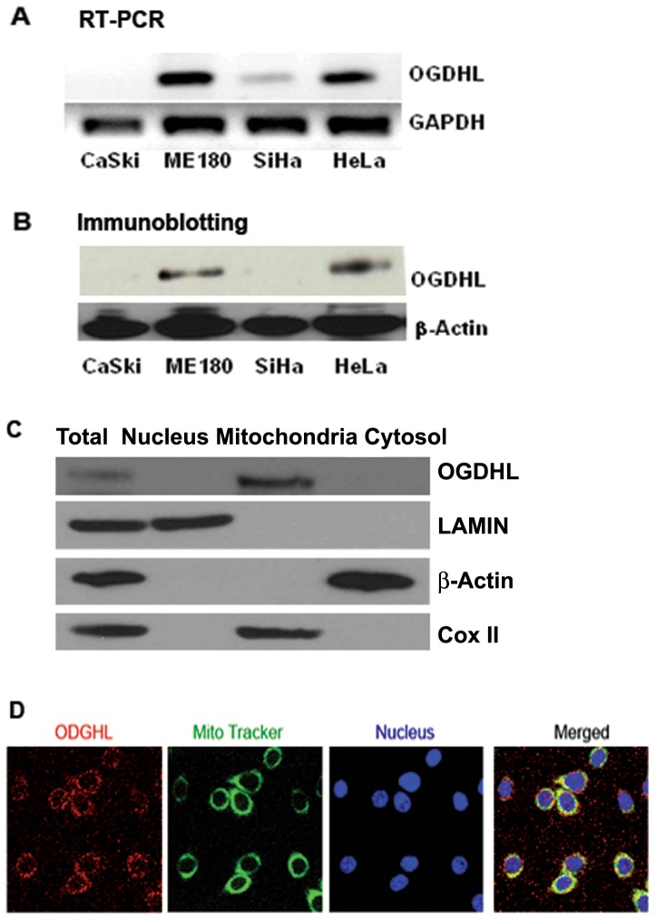
Figure 1. Expression and localization of OGDHL.
A. mRNA expression of OGDHL in different cervical cancer cell lines. B. Protein expression of OGDHL in different cervical cancer cell lines C. OGDHL, β-actin, lamin and Cox-II in total, nuclear, mitochondria and cytosolic fractions of ME180 cell lines. D. Colocalization of OGDHL and MitoTracker Green in ME180 cell line. Cultures were co-loaded with the OGDHL (red fluorescence) and the mitochondrial marker, MitoTracker Green (green fluorescence), and imaged with confocal microscopy. Note the substantial overlap between OGDHL and MitiTacker (yellow), indicating that they largely target the same intracellular organelles.

