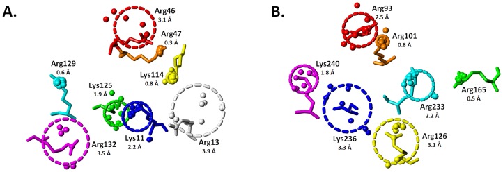
Figure 2. Radius (R g) of gyration for HBS basic residues:
the HBSs of the pentasaccharide binding sites of (A) antithrombin and (B) exosite II of thrombin are depicted with gyrational mobility as thick dashed lines that convey the circumference of movement. The radius of gyration (Å) is listed below each basic residue. The basic side chains from (A) 1TB6 and (B) the AB subunits of 1XMN are shown. See text for details.

