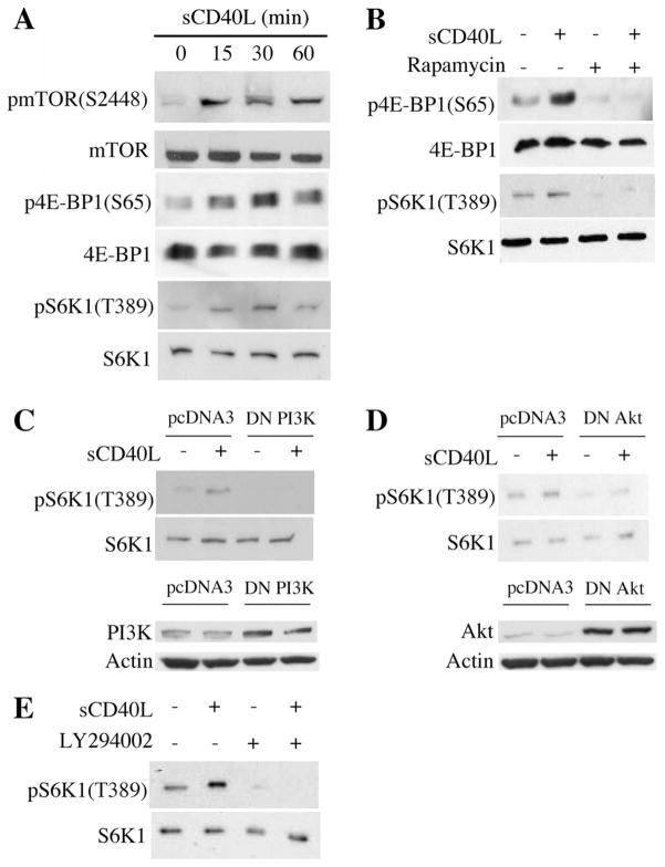
FIGURE 1.
Ligation of CD40 on EC activates mTOR and mediates mTORC1 signaling. A, Confluent cultures of EC were serum starved over-night and treated with 3 μg/ml sCD40L for the indicated times. Cells lysates were prepared and Western blotting was performed for total and phosphorylated mTOR, 4E-BP1, and S6K1. B, Confluent cultures of EC were serum starved overnight and treated with 10 ng/ml rapamycin 1 h before stimulation with 3 μg/ml sCD40L for 30 min. Western blotting analysis was performed using Abs to total and phophorylated 4E-BP1 and S6K1. C, upper panel, EC were transfected with a dominant negative mutant of PI3K (DN PI3K, 100 ng) or the empty vector (pcDNA3) as a control. After 48 h, the cells were serum starved overnight and treated with sCD40L (3 μg/ml) for 30 min. A Western blot illustrating total and phosphorylated S6K1 is shown. Lower panel, Protein expression of PI3K and Actin in EC following transfection with control pcDNA3 or the DN PI3K, evaluated by Western blot. D, EC were transfected with a dominant negative mutant of Akt (DN Akt, 100 ng) or pcDNA3 as a control and were treated with sCD40L as in C. A Western blot was performed to evaluate the expression of total and phosphorylated S6K1. Lower panel, The expression of Akt and Actin were evaluated by Western blot in empty vector (pcDNA3) and DN Akt-transfected EC. E, Serum starved EC were cultured with the PI3K inhibitor LY294002 (20 μM) for 1 h and stimulated with sCD40L (3 μg/ml) for 30 min. A Western blot of total and phosphorylated S6K1 was performed. The illustrated blots are representative of at least three experiments, all with similar results.

