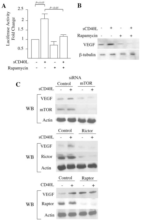
FIGURE 3.
mTORC2 regulates CD40-inducible expression of VEGF in EC. A, EC were transfected with a full-length 2.6-kb VEGF promoter-luciferase construct. Twenty-four hours following transfection, the cells were stimulated with sCD40L (3 μg/ml) in the absence or presence of rapamycin (10 ng/ml) for an additional 24 h. The cells were lysed and promoter activity was calculated as the fold change in luciferase counts from each group of cells, compared with untreated cells. Illustrated are the mean results of three independent experiments (± 1 SD). p values were calculated using the Student’s t test. Panel B, EC were cultured for 12 h in the absence or presence of rapamycin (10 ng/ml) before stimulation with sCD40L (3 μg/ml) for an additional 24 h. The cells were lysed and the expression of VEGF protein was analyzed by Western blot. The illustrated blot is representative of three experiments with similar results. The expression of β-tubulin served as an internal control. C, EC were transfected with control, mTOR, rictor, or raptor siRNA, and after 24 h, were cultured in the absence or presence of sCD40L (3 μg/ml) for an additional 24 h. Subsequently, the cells were lysed and the expression of VEGF was examined by Western blot (labeled WB). As a control for siRNA transfection and knockdown, each lysate was also simultaneously evaluated for the expression of either mTOR, rictor or raptor by Western blot, as indicated. The expression of actin served as an internal control. Each blot is representative of four with similar results.

