FIGURE 5.

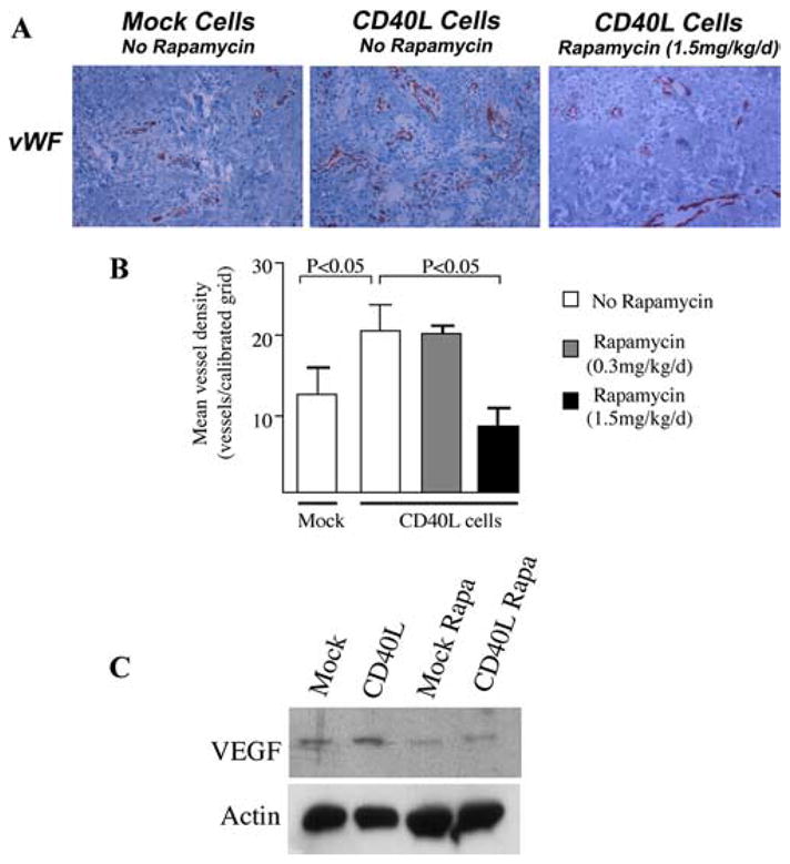
Function of mTOR in CD40L-mediated angiogenesis in vivo. SCID mice bearing human skin transplants received intracutaneous injections of mock transfectant cells (Mock Cells) or CD40L-expressing murine fibroblasts (CD40L Cells), and were left untreated or were treated with rapamycin (0.3 mg/kg/day or 1.5 mg/kg/day) by i.p injection. The human skin was harvested after 7 days and evaluated for angiogenesis by immunohistochemical staining for vWF (A and B), and for VEGF expression by Western blot (C). A, Representative photomicrographs of vWF staining of endothelial cells in Mock- or CD40L-injected skins (magnification ×400) from untreated or rapamycin treated mice. B, Mean vessel density (± 1 SD) in skins following injection of Mock cells (n = 5), CD40L cells in untreated animals (n = 11), or in animals treated with rapamycin (0.3 mg/kg/d, n = 10) or rapamycin (1.5 mg/kg/d, n = 8). p values were calculated using the Mann-Whitney U test. C, Representative Western blot of VEGF expression in Mock- and CD40L-injected skins harvested from mice untreated or treated with rapamycin (Rapa, 1.5 mg/kg/d). Representative of three with similar results.
