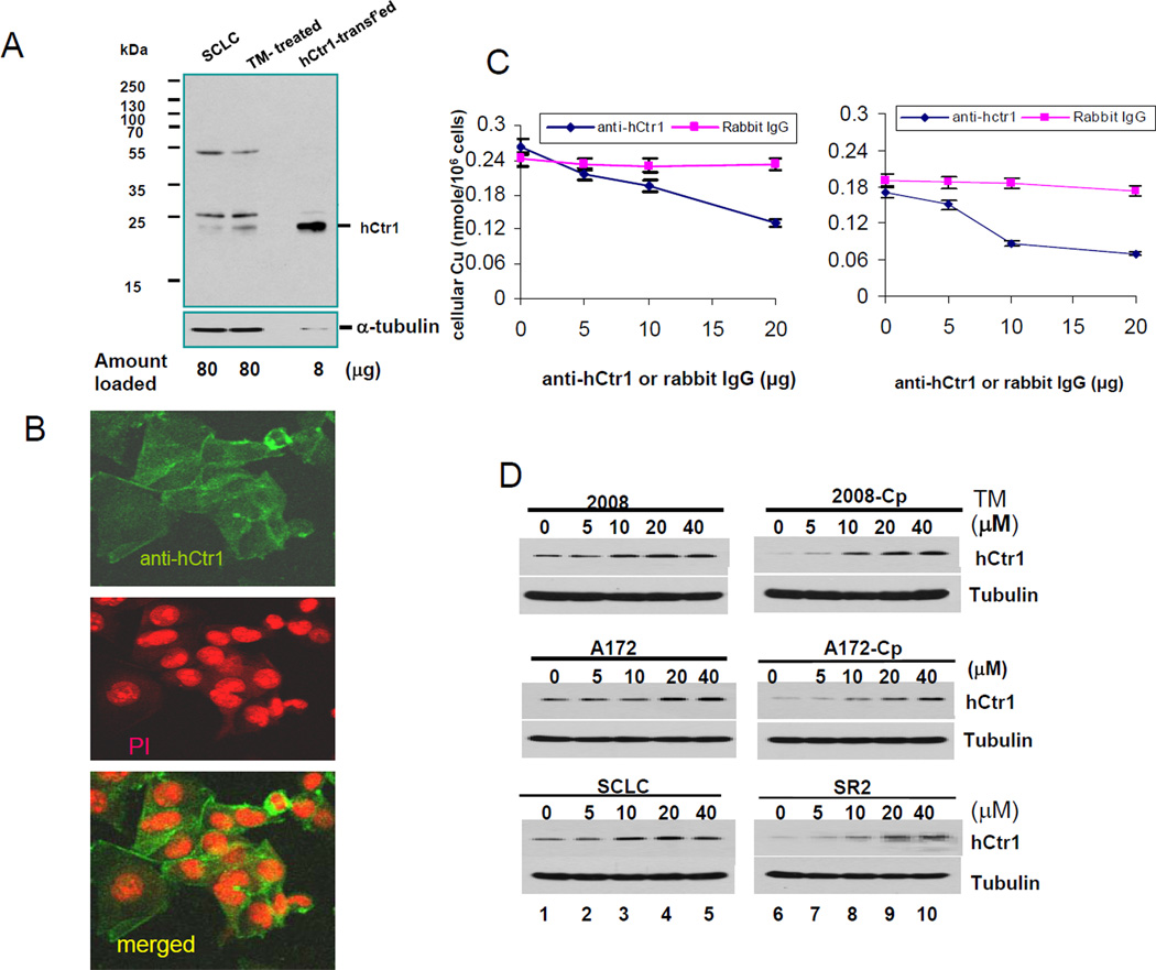
Fig. 3.
Characterization of anti-hCtr1 antibody. (A) Western blotting of hCtr1 expression in cell extracts prepared from different cells as indicated. The hCtr1-transfected sample was purposely under-loaded (0.1x the amount). (B) Confocal immunofluorescence microscopy of hCtr1 staining by anti-hCtr1 antibody (upper), PI staining (middle) and merged (lower). (C) Functional demonstration of the concentration-dependent suppression of Cu (left) and cDDP (right) transport by the hCtr1 antibody. (D) Western blotting determinations of hCtr1 expression in three pairs of cDDPR vs their corresponding sensitive cells treated with various concentrations of TM as indicated for 16 h.

