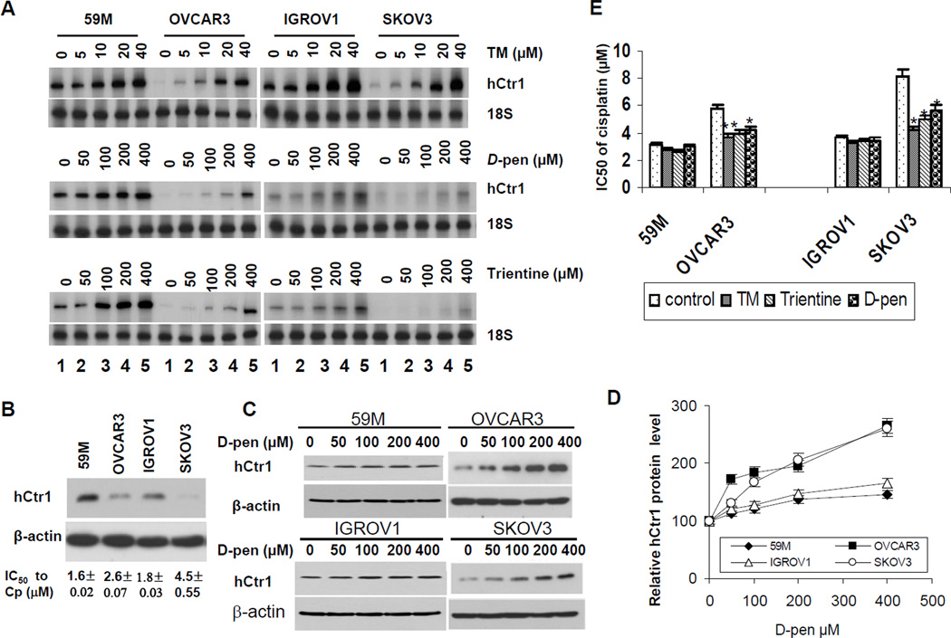
Fig. 4.
hCtr1 expression levels and re-sensitization to cDDP in four ovarian cancer cell lines treated with Cu-lowering agents. (A) RPA of hCtr1 mRNA levels in cells treated with different concentrations of TM (upper), D-pen (middle), and trientine (lower). (B) Western blotting analysis of hCtr1 expression (top) and sensitivity of these cell lines to cDDP (IC50 values shown in bottom). (C) Western blotting of ovarian cancer cell lines treated with D-Pen as indicated using β-actin as loading control. (D) Densitometric analyses of hCtr1 expression blotting results as shown in (C). (E) Sensitivity of ovarian cancer cells to cDDP in the presence and absence of Cu-lowering agents as indicated.

