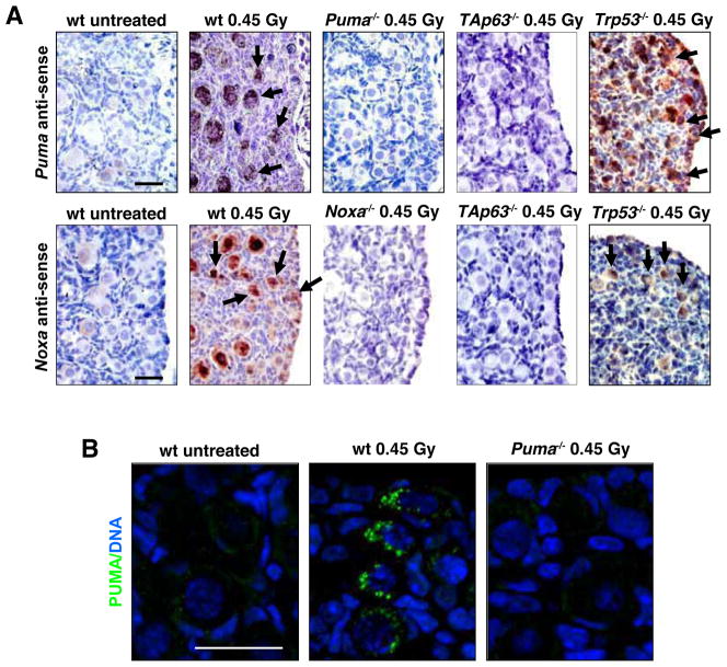
Figure 1. Expression of Puma mRNA, Noxa mRNA and PUMA Protein in Primordial Follicle Oocytes Following γ-irradiation-induced DNA damage.
(A) Ovaries were harvested from PN5 wt, Puma−/− (negative control), Noxa−/− (negative control), Trp53−/− and TAp63 mutant mice at 0 (untreated control) and 3 h post whole-body γ-irradiation (0.45 Gy). In situ hybridization (top panel) was performed using anti-sense probes for Puma and Noxa. Puma and Noxa mRNAs were detected in primordial follicle oocytes from wt and Trp53−/− mice but not in those from TAp63 mutant mice, 3 h post γ-irradiation. Arrows indicate positively stained primordial follicle oocytes (dark purple/brown staining). Control experiments, including in situ hybridization of sections from Puma−/− ovaries with Puma anti-sense probes and in situ hybridization of sections from Noxa−/− ovaries with Noxa anti-sense probes are shown in Figure S1. (B) PUMA antibody immunofluorescent staining (bottom panel; green) in wt and Puma−/− (negative control) primordial follicle oocytes at 6 h post γ-irradiation. Scale bar: 20 μm.

