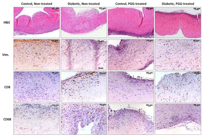
Figure 4.
Cell infiltration in collagen scaffolds. Decellularized porcine aortic valve cusps treated with PGG or non-treated controls were implanted subdermally in control and diabetic rats. Explants were stained by Hematoxylin and Eosin (H&E, dark purple=nuclei, pink=background substance) and immunohistochemistry for vimentin (Vim.), CD8 (T-lymphocytes) and CD68 (macrophages). Positive IHC reaction=brown.

