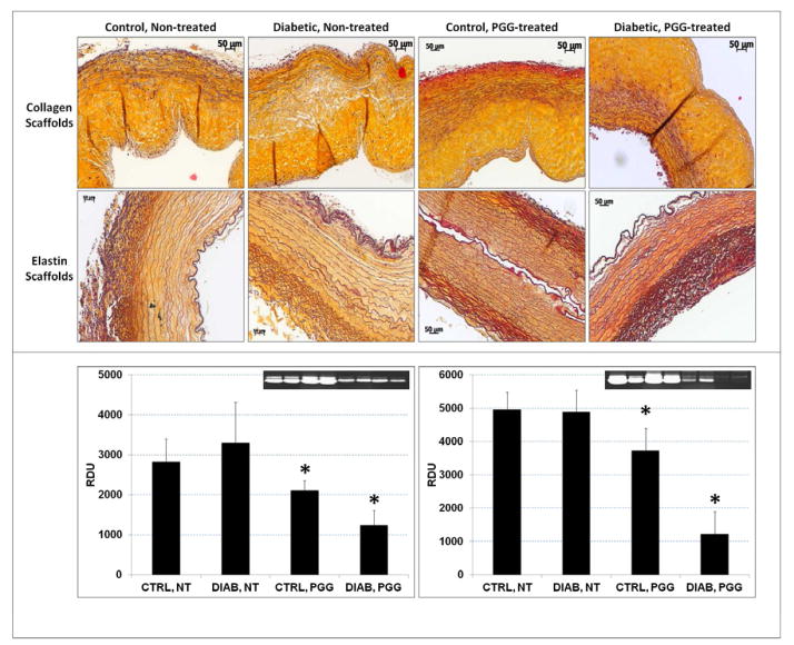
Figure 6.
ECM remodeling in implanted scaffolds. Scaffolds treated with PGG or non-treated controls were implanted subdermally in control and diabetic rats. (upper panel) Explants were stained with Movat’s Pentachrome histology stain (yellow=collagen, blue=glycosaminoglycans, dark purple=elastin, bright red=nuclei). (bottom panel) Protein extracts from collagen scaffolds (left) and elastin scaffolds (right) were analyzed for matrix metalloproteinase activity by gelatin zymography followed by densitometry (inserts, positive=white bands); results are shown as relative density units (RDU). *indicates statistical significance.

