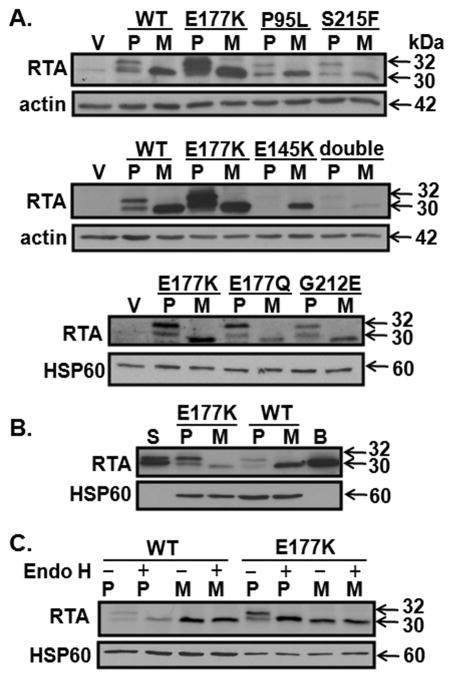
Figure 2.
Expression of RTA and RTA mutants in transfected MAC-T cells. Total cell lysates (A. 50 μg per lane; B. 50 μg per lane for vector and wt; 25 μg for E177K) were collected from MAC-T cells 19 h after transfection. Membranes were immunoblotted with anti-RTA antibody then stripped and reprobed with antibodies to actin or HSP60. Blots are representative of 2 to 3 experiments. S = RTA glycosylated standard from Sigma (5 ng); B = non-glycosylated RTA standard from BEI (10 ng). C. Total cell lysates (40 μg for WT and 20 μg for E177K) were treated with Endo H prior to immunoblotting. V = vector; P = pre; M = mature; WT = wild-type; double = P95L/E145K.

