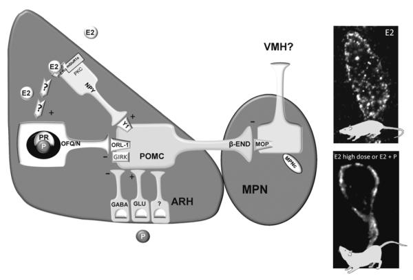
Figure 1.
Proposed model of estradiol regulation of lordosis circuitry within the ARH and medial preoptic nucleus (MPN). Estradiol binds to that signals through PKC to induce the release of neuropeptide Y (NPY; although the mERα-mGluR1a is depicted in the NPY neuron it may located in a neuron upstream of the NPY neuron). NPY then binds to the NPY-Y1 receptor (Y1) to excite (+) a subpopulation of POMC neurons involved in sexual behavior that project to the MPN. Estradiol rapidly activates this circuit and maintains activation of POMC neurons to induce the release of β-END from nerve terminals in the MPN. β-END binds to and activates and internalizes μ-opioid receptors (MOP) into early endosome to initially inhibit lordosis (E2 photomicrograph). A priming dose of estradiol maintains MOP activation for at least 30 hours inhibiting lordosis and increases ORL-1 expression in ARH POMC neurons that project to the MPN. This circuit is deactivated by longer (48 hours) exposure to high doses of estradiol (5-50 μg; E2 high dose photomicrograph) by 1) reducing NPY release through possibly the downregulation of mERα-mGluR1a in NPY neurons and 2) through an unknown mechanism increasing the release of OFQ/N to activate ORL-1 which decreases POMC excitation (-) through increasing GIRK currents. This reduces MPN MOP activation, recycling the MOP to the plasma membrane as part of the mechanism for facilitating lordosis. Following estradiol priming, progesterone also deactivates this circuit to facilitate lordosis. Although progesterone receptors are expressed in OFQ/N neuron, OFQ/N is not necessary for deactivating MPN MOP and progesterone may induce the release of other inhibitory neurotransmitters such as GABA to inhibit β-END release to facilitate lordosis [49, 85, 86, 102, 144, 248, 339, 340, 358, 362, 416].

