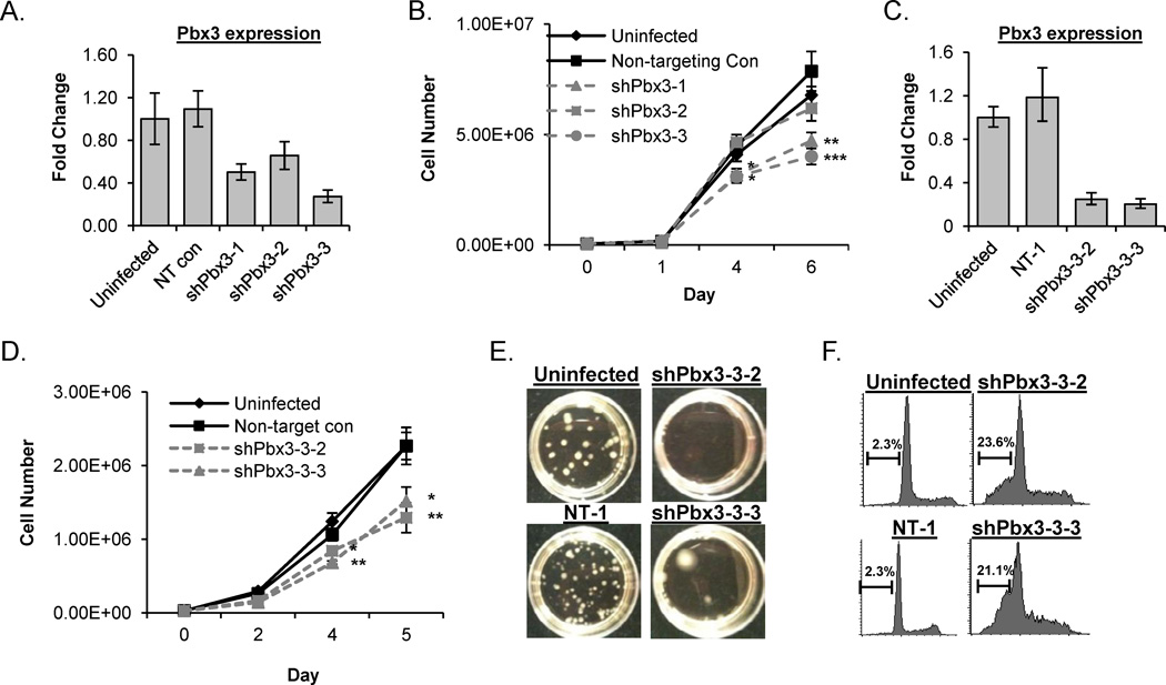
Figure 5. Knockdown of Pbx3 inhibits proliferation and promotes apoptosis in a NHD13 cell line.
(A) RQ-PCR analysis for Pbx3 expression from pools of 189MIG cells infected with shRNA against a non-targeting (NT) control or Pbx3 (shPbx3-1, shPbx3-2, and shPbx3-3) compared to uninfected 189 MIG cells. (B) Proliferation curves of pooled clones from uninfected, NT control, shPbx3-1, shPbx3-2, and shPbx3-3 infected cells. (C) RQ-PCR analysis of Pbx3 expression from single cell clones of 189MIG cells infected with the NT control (NT-1) and two clones infected with shPbx3-3 (designated shPbx3-3-2 and shPbx3-3-3), compared to uninfected 189MIG cells. (D) Proliferation curves from single cell clones from NT-1 and shPbx3-3-2 and shPbx3-3-3 compared to uninfected 189MIG cells. (E) Analysis of uninfected, NT-1, shPbx3-3-2 and shPbx3-3-3 in methylcellulose CFU assay (F) Measurement of DNA content from uninfected 189MIG, NT-1 shRNA control, shPbx3-3-2 and shPbx3-3-3 at day 5 in liquid culture. Apoptotic events are those with less than 2N DNA. * , **, and *** indicate p<0.05, 0.01, and 0.001 respectively.

