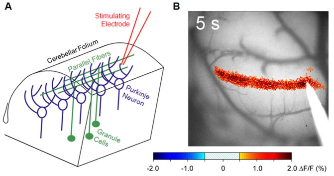
Figure 1.
Optical imaging of the parallel fiber-Purkinje neuron synapse. (A) Schematic diagram of cerebellar anatomy. The stimulating electrode is placed on the surface of the cerebellum after removal of the skull to activate multiple parallel fibers, leading to activation of Purkinje cells aligned in a mediolateral orientation. (B) Change in flavoprotein fluorescence within Cerebellar Purkinje neurons following parallel fiber activation. Changes in fluorescence are directly due to glutamatergic neurotransmission between the parallel fiber and the Purkinje neuron. Due to the circuitry of the cerebellum, electrode stimulation results in a “beam” of synaptic activity.

