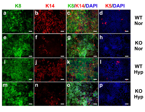
Figure 2.
Loss of basal marker expression in cultured HIF-1 knockout cells. Wild-type (WT) or knockout (KO) cells were plated onto tissue-culture-treated slides and grown to subconfluence, at which time subsets of cells were exposed overnight to hypoxic (Hyp) culture or remained under normoxic (Nor) culture. Cells were costained with Troma-I (K8, green) and K14 (red) (a) to (c), (e) to (g), (i) to (k) and (m) to (o) or with K5 alone (d), (h), (l) and (p); images showing either the green (K8) or red (K14) channel from the co-stained slides are also presented (a) to (b), (e) to (f), (i) to (j) and (m) to (n). All slides were counterstained with 4',6-diamidino-2-phenylindole (DAPI). Images were captured at ×200 original magnification (scale bar = 50 μm).

