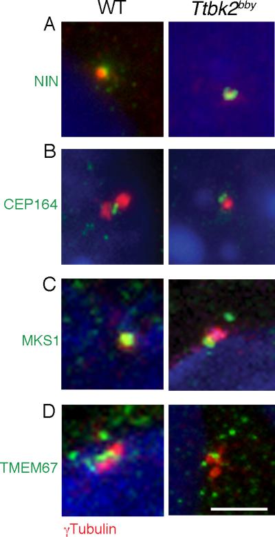
Figure 3. Normal localization of appendage and transition zone markers in Ttbk2bby basal bodies.

(A) The subdistal appendage marker Ninein (NIN; green) is expressed normally in Ttbk2bby MEFs. γ-tubulin (red) labels the centrioles. Images shown are representative of 14 cells imaged for Ttbk2bby and 12 for wild-type. (B) Localization of the distal appendage-associated protein CEP164 (green) distal to the centrosome, marked by γ-tubulin (red) in MEFs derived from WT and Ttbk2bby mutant embryos. 23 cells were imaged for Ttbk2bby and 20 for wild-type. (C, D) Transition zone proteins MKS1 (C) and TMEM67 (D) (green) are expressed in the transition zone of WT and at the distal basal body in Ttbk2bby cells. Centrosomes are labeled by γ-tubulin (red). Scale bar = 2 μm. Total number of cells imaged for each marker was: MKS1- 17 for Ttbk2bby; 13 for wild-type; TMEM67- 19 for Ttbk2bby; 22 for wild-type. Similar images were obtained from a minimum of 2 independent experiments. Please see Figure S3 for additional images.
