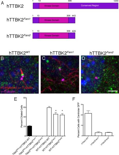
Figure 7. SCA11-associated variants of hTTBK2 do not rescue cilia formation in Ttbk2bby cells and interfere with ciliogenesis in WT cells.
(A) Schematic of human TTBK2, which is 89% identical to mouse TTBK2; and two familial SCA11 mutations that truncate the protein at 443 or 428 AA. (B-D) Localization of WT hTTBK2::eGFP (A) and SCA11-associated Fam1 and Fam2 (C and D) variants expressed in Ttbk2bby MEFs. GFP is in green, γ-tubulin is in purple, and acetylated α-tubulin is in red. The Fam1 and Fam2 fusion proteins generally localized diffusely in the cytoplasm; C show a rare cell in which GFP is localized to centrosome. Scale bar = 2 μm. (E) Graph showing the percentage of ciliated cells observed in Ttbk2bby mutant (black bars) or WT (white bars) MEFs infected with the indicated construct. (F) Graph showing the percentage of cells with centrosomal localization of GFP for the indicated construct. For E and F, bars represent the mean percentage of ciliated cells (E) or cells with centriolar GFP (F) from 4-8 randomly selected fields of cells per condition. Fields were selected from at least two different slides and had a minimum of 25 cells per field; see Supplemental Methods. Error bars indicate the SEM. Please see also Figure S6.

